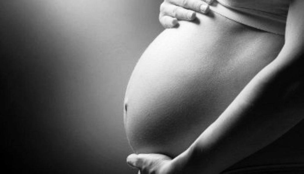
In 2002 this office was created with high sense of responsibility for the problems that may be encountered the woman at all ages. The knowledge, the international continuing education and the high technology equipment help in providing new and minimal invasive techniques equally effective in areas of rapid development. The collaboration and continuous audit of the Fetal Medicine Foundation in London by Professor Kypros H. Nicolaides guarantees high specialization in the field of maternal-fetal medicine and make us pioneers in the area. Working with scientists specialized in the field of genetics, embryology, hematology, child, adolescent and adult endocrinology, dysmorphology and plastic surgery enable organized treatment in many problems. The effort is continuing with responsibility, seriousness and experience.
Obstetric
Gynecology
Ultrasound
Colposcopy
see services
see services
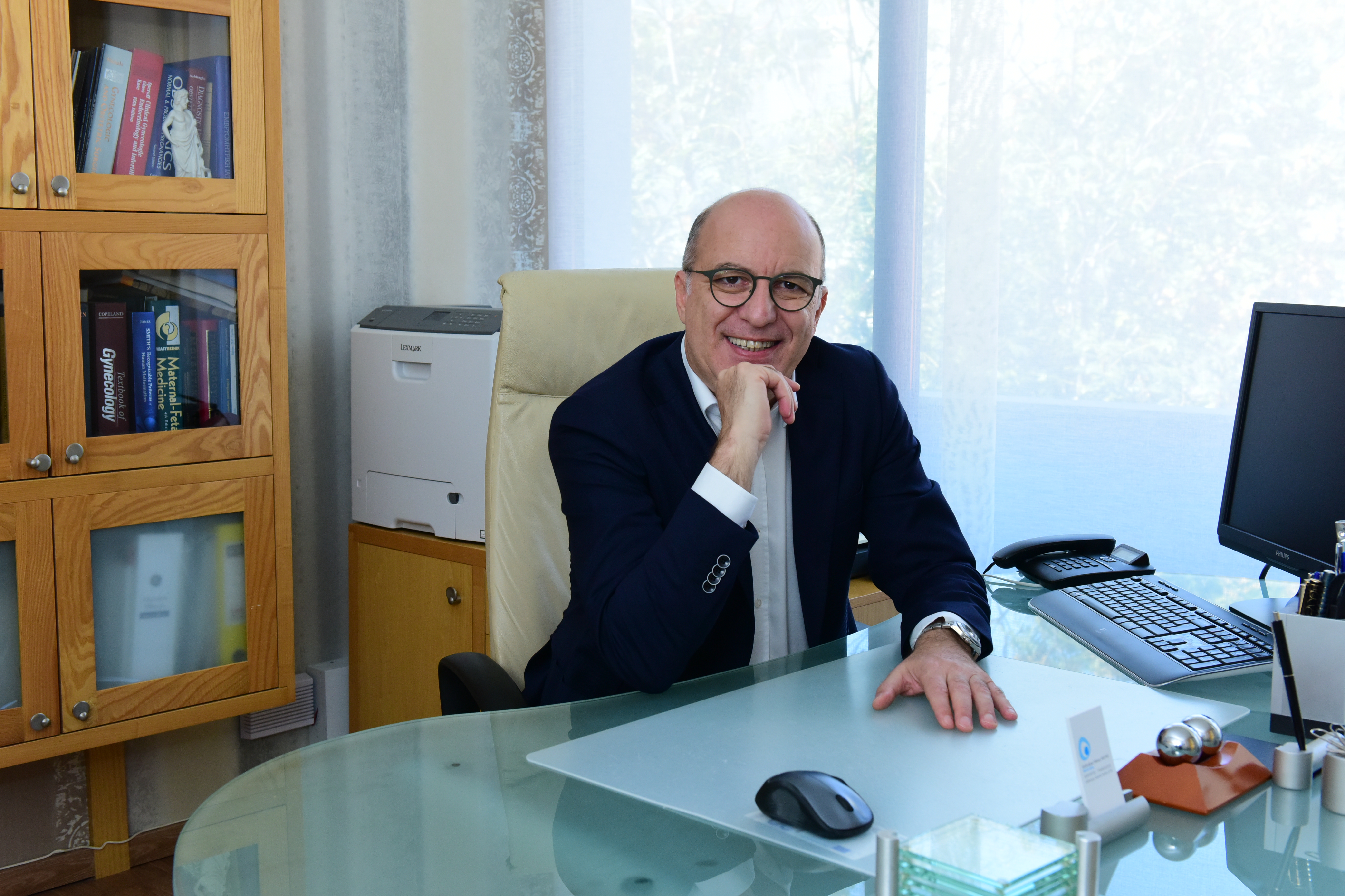
Head doctor:
Alexandros Mainas MD Ph.D
Midwife responsible for coordination:
Asimina Avzoti University of Thessaloniki
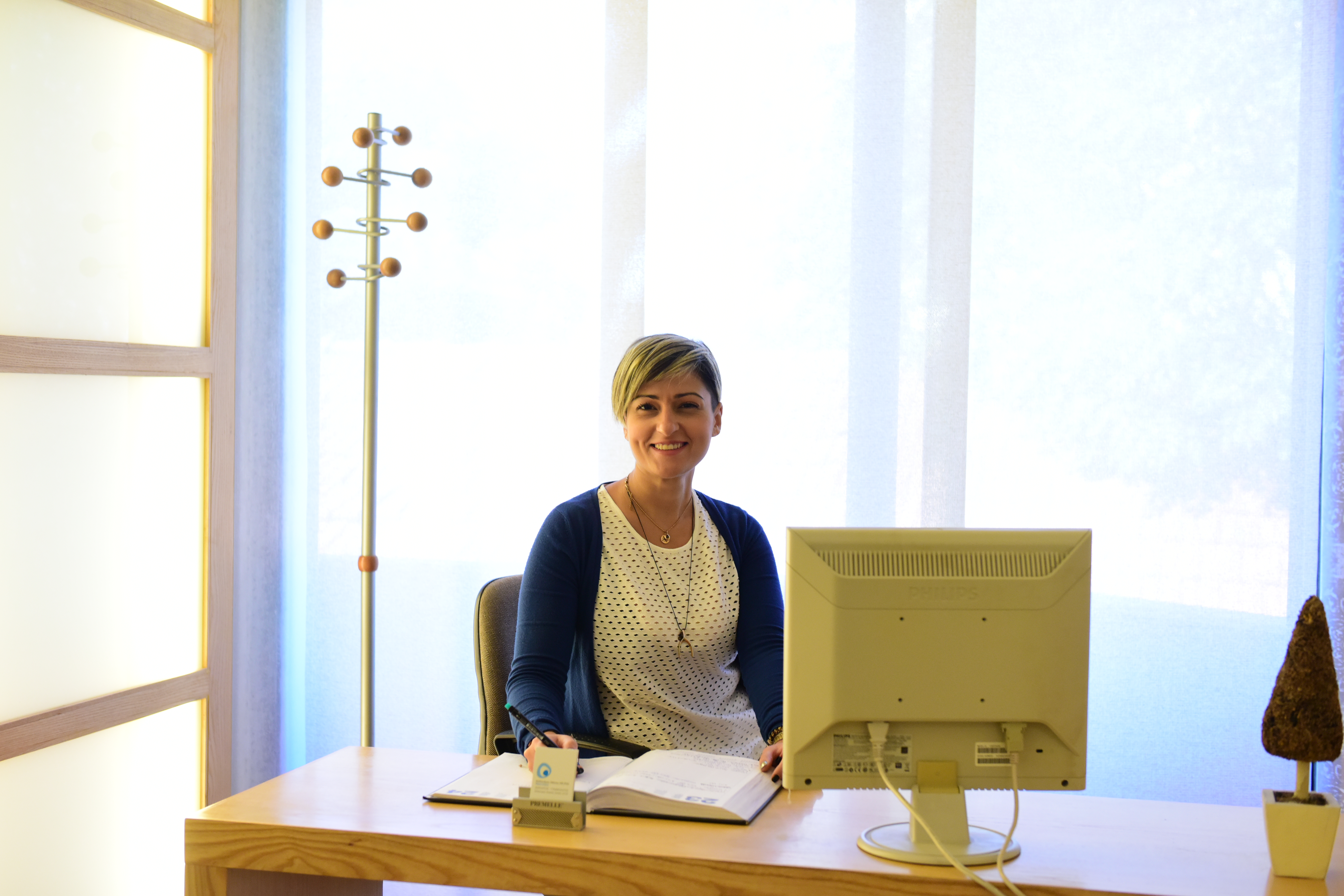
Administrative staff:
Eleni Papadopoulou
Time of services only by appointment
Daily from 09.00 to 13.00 & 18.00 to 22.00
except Wednesday and Saturday afternoon.
Short CV
1992 I graduated from the Medical School of Democritus University of Thrace among the first graduates of the year.
2001 I received the title of Specialization in Obstetrics - Gynecology, from University Hospital of Patras Greece
2002 I certified by the Fetal Medicine Foundation of London, UK to perform full antenatal ultrasound scan of the fetus.
2003 official license to perform ultrasound by the Greek Minister of Health.
2007 I received my Doctor of Philosophy degree (Ph.D) from the Medical School of Democritus University of Thrace with a grade "excellent" in a thesis with the title " Permeability of the placenta"
2015 certified by the Hellenic Society for Ultarsound in Obstetrics and Gynecology in prenatal diagnosis
I have participated in many conferences with free communications while my papers published in recognized journals. I am fluent in English and in Italian.
Advanced imaging technology
1. Ultrasound nuchal translucency
The most common trisomies are those of chromosomes 21, 18 and 13. Trisomy 21 is found in about 1 in 700 births and the risk increases with maternal age. The condition is associated with intellectual disabilities and some physical defects and most commonly heart abnormalities. The life expectancy is about 60 years. Trisomies 18 and 13 are found in about 1 in 7,000 births and the risk increases with maternal age. The conditions are associated with severe mental handicap and several physical defects. Most affected individuals die before or soon after birth and they rarely survive beyond the first year of life.
Today we assess the risks of Down's syndrome and other chromosomal abnormalities. Each woman will be given an estimate of her individual risk for this pregnancy. This is calculated by taking into account the age of the mother, measurement of two hormones in the mother’s blood and the scan findings of nuchal translucency thickness, nasal bone , and blood flow through the tricuspid valve of the fetal heart and ductus venosus and fetal abnormalities. In the Greek version you can find a video by You tube of the nuchal scan. Parents will receive full counseling concerning the significance of these risks and the various options for further investigations including invasive testing or the cfDNA test. In addition the aim of the nuchal scan is:
To diagnose multiple pregnancy. Approximately 2% of natural conceptions and 10% of assisted conceptions result in multiple pregnancy. Ultrasound scanning can determine if both babies are developing normally and if the babies share the same placenta which can lead to problems in the pregnancy. In such cases it would be advisable to monitor the pregnancy more closely.
To diagnose major fetal abnormalities. Some major abnormalities may be visible at this gestation. However it will still be necessary to have a 20 week anomaly scan.
To diagnose early miscarriage. Unfortunately, in 2% of women who attend for a nuchal scan it is found that the fetus has died, often several weeks before and without any warning. Couples will receive full counseling as to the possible causes of this problem and the options for subsequent measures that may be necessary.
To estimate the probability of early preclampsia, intrauterine retardation of the fetus, and spontaneous premature birth (FMF certified).
2. Detailed Anatomical Survey Ultrasound
3. Doppler ultrasound scan (Certified by Fetal Medicine Foundation of London)
Doppler assessment of the fetal circulation plays an important role in assessment of the fetal condition in pathological pregnancies. With this scan we assess the flow of the blood in the main arteries and veins of the fetus.
4. 4D fetal ultrasound
Maternal-fetal medicine
Monitoring of low and high risk pregnancies (diabetes, control-preventing preterm delivery, hypertension, thyroid disorders, maternal thrombophilic disorders and multiple pregnancies, etc.)
Genetic counseling in collaboration with a clinical geneticist and genetic specialist in Dysmorphology for: Congenital anomalies of fetus, babies, and children, hereditary diseases, Ethnicity Susceptible Concerns, counseling for family history, consanguinity, exposure to teratogenic drugs, unexplained miscarriages, mental retardation, autism, etc.
Invasive and non invasive prenatal diagnosis
Amniocentesis
Non Invasive Prenatal Testing (NIPT)
Maternal Serum Screening
Non-Stress Testing (NST)
Genetic tests (molecular, biochemical , cytogenetics) in collaboration with ATG Genome
Gynecology
Check-up (gynecological examination, Pap test, transvaginal ultrasound, breast examination)
Sono-hysterography
(uterine endometrial polyps, fibroids, abnormal bleeding)
Procedures without anesthesia
(Pipelle endometrial biopsy, insertion IUD, etc.)
Colposcopy, cervical biopsies
Prevention of gynecological cancer
Subfertility- Services of assisted reproduction
Investigation of the infertile couple
Induction of ovulation
Intrauterine insemination
Application protocols of assisted reproduction, egg and sperm donation, and embryos cryoreserved for natural or surrogate mothers.